- Home
-
Products
- Diamond Burs For Clinic
- Tungsten Carbide Burs
- Surgical Tungsten Carbide Burs
- Triming Finishing Gold Tc Bur
- Diamond Stones & Diamond Polishers
- Diamond Rubber Polishers
- Cleaning Burs After Debonding
- Silicon Polishers
- White Stones & Green Stones
- Polishing Brushes
- Mandrels
- Blocks, Sterilisable
- Clinic Kits
- Samples Collection
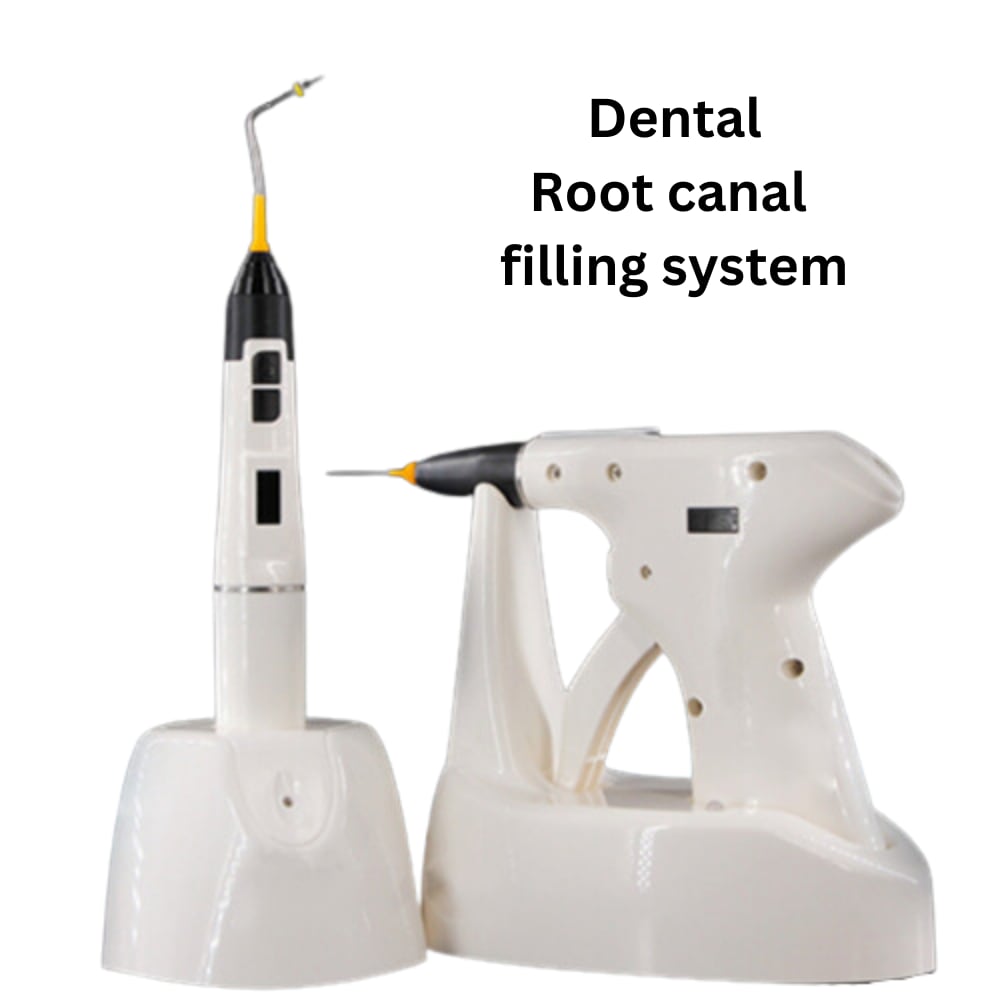
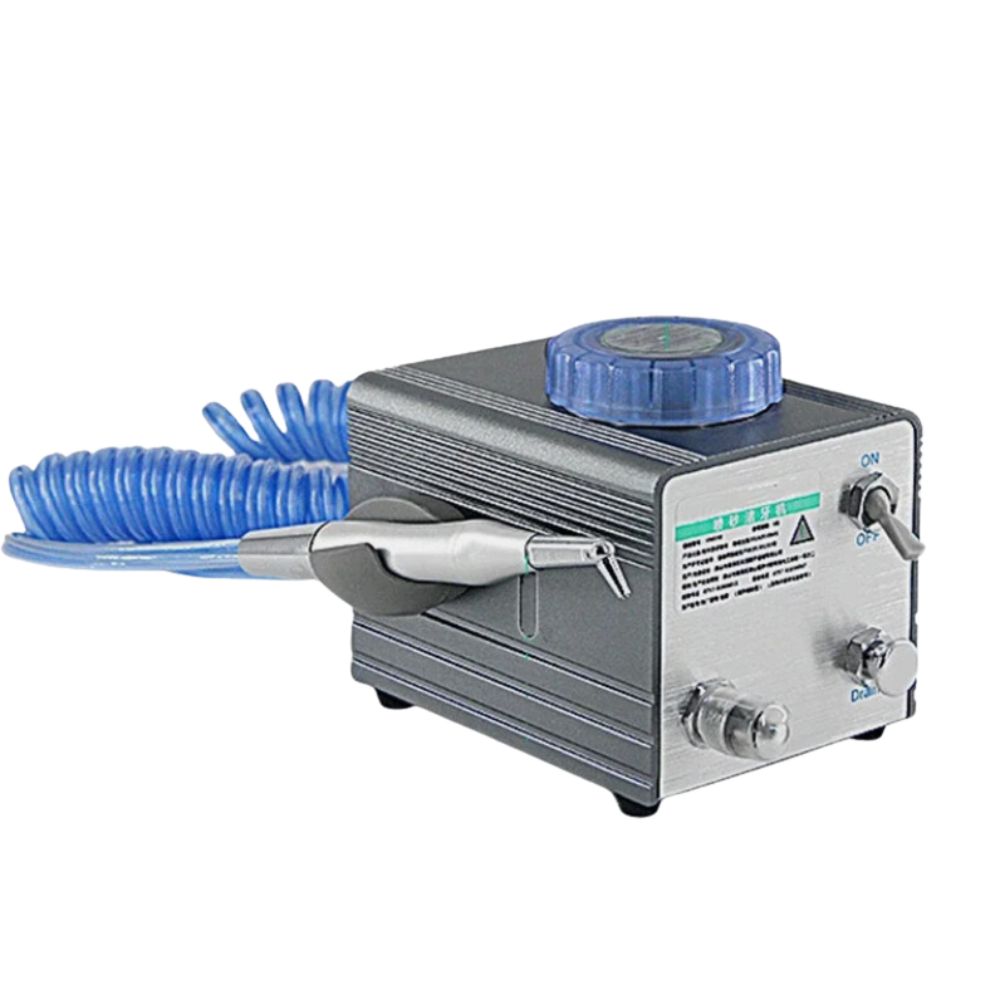
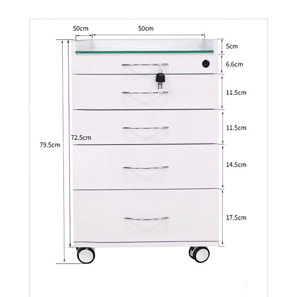
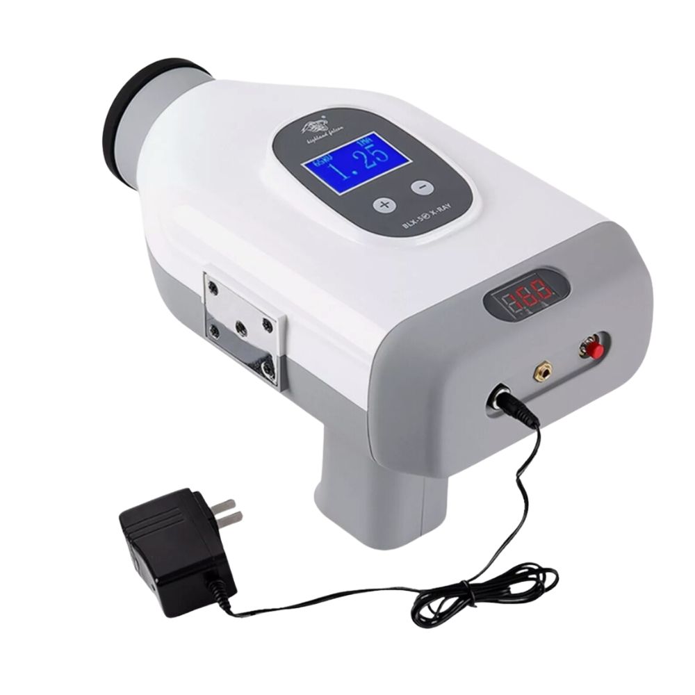
- Dental Equipment
- Dental Handpiece & Implant Bender
- Dental Mobile Photography
- Diamond Bur Hp
- Fully Sintered Diamond Bur Hp
- Diamond Disc
- Plaster Trimming Wheel
- Cad/cam Milling Burs
- Tungsten Carbide Bur
- Tungsten Carbide Cutter Hp
- Tungsten Carbide Burs For Milling Machines
- Diamond Stones For Zirconia Or All Ceramics
- Diamond Polishers For Zirconia Or All Ceramics
- Silicon Polisher
- Silicon Polishing Burs For Composite Resins
- Mounted Stone
- Mounted Sandpaper
- Polishing Brushes & Felt Bob & Burmandrels
- Polishing Brush & Wheel In Bristle Cotton Cloth
- Felt Bobs & Burs
- Hard Z Cutter
- Sharpening Stones
- Cleaning Brushes By Hand
- Stainless Steel Discs
- Stone Disc
- Technician Kits
- Flexsoft Partial Denture
- Empty Aluminum Cartridge
- Twin-Color Diamond polishers
- Dental Porcelain Brushes
- About Us
- Blog
- Catalogs
- Contact Us