Digital Dental X-ray Sensors: Complete Benefits Guide
Digital radiography has transformed modern dental practices, replacing traditional film-based systems with advanced sensor technology. Digital dental X-ray sensors deliver immediate, high-resolution images while reducing radiation exposure and operational costs. Understanding the different sensor types and their benefits helps dental professionals make informed equipment decisions.
Digital vs. Traditional Film Radiography
The transition from film to digital radiography represents one of the most significant advancements in dental imaging. Traditional film required chemical processing, darkroom facilities, and waiting periods before images could be reviewed. Digital sensors eliminate these delays, displaying images on screen within seconds of exposure.
Film radiography presented several inherent limitations. Chemical processing required careful temperature control and fresh solutions to maintain image quality. Storage of physical films consumed significant space, and retrieval of archived images could be time-consuming. Environmental concerns also arose from chemical waste disposal.
Digital systems address these challenges while providing additional diagnostic capabilities. Image enhancement tools allow practitioners to adjust contrast, brightness, and magnification without retaking exposures. This flexibility improves diagnostic accuracy and reduces patient radiation exposure from retakes.
Types of Digital Dental X-ray Sensors
CCD Sensors (Charge-Coupled Device)
CCD sensors were among the first digital radiography technologies adopted in dentistry. These sensors use a silicon chip divided into pixels that convert X-ray photons into electrical charges. The charges are then processed into digital images.
CCD sensors typically feature rigid construction with fiber optic coupling between the scintillator layer and the CCD chip. This design provides excellent image quality with high resolution and contrast. However, the rigid sensor body can present challenges with patient comfort, particularly for posterior radiographs or pediatric patients.
CMOS Sensors (Complementary Metal-Oxide Semiconductor)
CMOS technology represents the newer generation of digital sensors. Each pixel in a CMOS sensor contains its own amplifier, allowing for faster image processing and lower power consumption compared to CCD sensors.
Many manufacturers have developed thinner CMOS sensors with improved ergonomics. The reduced thickness enhances patient comfort during image capture. CMOS sensors also tend to generate less heat during operation and may offer longer service life due to their simplified electronic architecture.
PSP Plates (Photostimulable Phosphor)
PSP plates, also called storage phosphor systems, function differently from direct digital sensors. These flexible plates are similar in size and shape to traditional film packets, making them familiar to practitioners and more comfortable for patients.
After X-ray exposure, PSP plates must be scanned by a special reader device that uses laser light to release the stored energy as light, which is then converted to a digital image. While this process takes longer than direct digital sensors, PSP plates offer advantages in patient positioning flexibility and lower initial equipment costs.

Key Benefits of Digital Dental X-ray Sensors
Reduced Radiation Exposure
Patient safety remains paramount in dental radiography. Digital sensors require significantly less radiation to produce diagnostic-quality images compared to film. Studies demonstrate radiation reduction of 50-90% depending on the sensor type and imaging protocol used.
This substantial reduction in radiation dose benefits both patients and dental staff. For practices performing high volumes of radiographic imaging, cumulative radiation exposure reduction becomes particularly significant over time.
Instant Image Availability
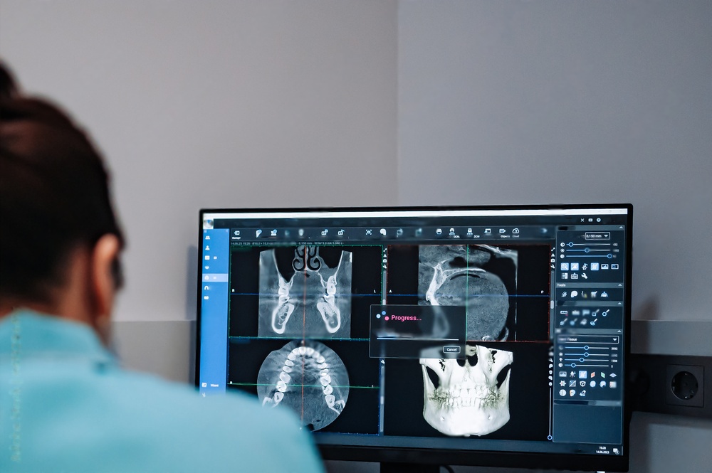
The elimination of chemical processing time transforms clinical workflow. Images appear on the computer screen within seconds, allowing immediate review during the patient appointment. This immediate feedback enables practitioners to verify proper image capture and positioning before the patient leaves the operatory.
Real-time image availability also enhances patient communication. Practitioners can display and discuss radiographic findings with patients immediately, improving case acceptance and treatment understanding. Educational software can overlay diagrams or annotations directly on the radiographic images.
Superior Image Quality and Enhancement
Digital sensors capture images with excellent resolution and dynamic range. The ability to manipulate images after capture provides diagnostic advantages not possible with film. Contrast adjustment helps visualize subtle differences in tissue density. Magnification tools allow detailed examination of specific areas without additional exposures.
Color enhancement and edge detection filters can highlight anatomical structures or pathology. Measurement tools enable precise calculation of distances, angles, and areas. Some systems include diagnostic aids that analyze bone density or identify potential carious lesions.
Efficient Image Storage and Retrieval
Digital images integrate seamlessly with practice management and patient record systems. Radiographs stored digitally require no physical space and suffer no degradation over time. Electronic storage facilitates compliance with record retention requirements while eliminating concerns about film deterioration.
Image retrieval becomes instantaneous with proper digital organization. Comparison of current and previous radiographs helps track treatment progress or disease progression. Images can be transmitted electronically to specialists, insurance companies, or referring practitioners without quality loss.
Cost Savings and Environmental Benefits
While digital sensors require higher initial investment than film holders, operational cost savings accumulate quickly. Elimination of film purchase, processing chemicals, and darkroom maintenance reduces ongoing expenses significantly.
Environmental benefits extend beyond cost savings. Digital radiography eliminates chemical waste disposal, reduces water consumption, and decreases the environmental impact of film manufacturing and packaging materials.
Selecting the Right Digital Sensor System
Several factors should guide sensor selection for your practice. Image quality requirements, patient comfort considerations, budget constraints, and integration with existing systems all play important roles in the decision process.
| Consideration | CCD Sensors | CMOS Sensors | PSP Plates |
|---|---|---|---|
| Image Quality | Excellent resolution | Excellent resolution | Good resolution |
| Patient Comfort | Moderate (rigid) | Good (thinner options) | Excellent (flexible) |
| Image Speed | Immediate | Immediate | Delayed (scanning required) |
| Initial Cost | High | Moderate to High | Lower |
| Durability | Good with care | Good with care | Plates require replacement |
Sensor size options should match your patient demographics. Pediatric practices may benefit from smaller sensor sizes, while practices treating primarily adult patients might prioritize larger sensors that capture more anatomy per exposure.
Software integration capabilities affect workflow efficiency. Ensure the sensor system interfaces properly with your practice management software, allowing seamless image transfer and storage within patient records.
Proper Sensor Maintenance and Care
Digital sensors represent significant investments that require proper care to maximize longevity and maintain image quality. Infection control procedures must balance thorough disinfection with protection of sensitive electronic components.
Infection Control Protocols
Barrier protection remains the primary infection control method for digital sensors. Disposable plastic sheaths prevent direct contact between sensors and oral tissues. Multiple barrier layers may be used for added protection, particularly for sensors with irregular shapes or connection points.
After barrier removal, sensor surfaces should be cleaned and disinfected according to manufacturer recommendations. Most sensors cannot withstand immersion or high-level disinfection, so surface disinfection with appropriate agents becomes necessary. Always verify that disinfectant products are compatible with sensor materials.
Cable and Connector Care
Cable damage represents a common cause of sensor failure. Avoid sharp bends, particularly near the sensor connection point. Cable strain relief should be maintained during use and storage. Never pull sensors by the cable or allow heavy objects to rest on cables.
Connection points require gentle handling. Forcing connectors or connecting sensors while the system is powered on can damage delicate electronics. Establish a standard connection sequence and train all staff members on proper procedures.
Storage and Handling
Dedicated sensor storage protects against physical damage when sensors are not in use. Wall-mounted holders or drawer organizers prevent cables from tangling and sensors from being knocked off counters. Store sensors in locations away from excessive heat, moisture, or chemical exposure.
Regular inspection identifies potential problems before they cause sensor failure. Check cables for fraying or damage, examine sensor housings for cracks, and test image quality periodically. Address any issues promptly to prevent more extensive damage.
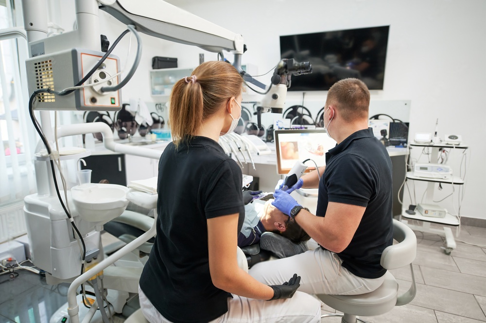
Integrating Digital Sensors with Dental Instrumentation
Modern dental practices combine digital imaging with advanced instrumentation for optimal patient care. High-quality diamond dental burs and tungsten carbide burs work alongside digital radiography to deliver precise diagnosis and treatment.
Radiographic imaging guides proper bur selection for cavity preparation or crown reduction. Digital measurements help determine appropriate bur sizes and shapes for specific procedures. The combination of accurate imaging and quality instrumentation improves treatment outcomes and efficiency.
Future Developments in Digital Dental Imaging
Sensor technology continues to advance with smaller form factors, higher resolution, and improved wireless connectivity. Artificial intelligence applications are being developed to assist with radiographic interpretation, identifying potential pathology or measuring anatomical structures automatically.
Integration between intraoral scanners and radiographic sensors may provide complete three-dimensional diagnostic information. These developments promise to further enhance diagnostic capabilities while maintaining or reducing radiation exposure levels.
Making the Transition to Digital Radiography
Practices still using film radiography should evaluate the benefits of transitioning to digital sensors. The learning curve for digital radiography is relatively short, with most practitioners becoming proficient within weeks of implementation.
Staff training should cover sensor positioning, infection control procedures, software operation, and troubleshooting common issues. Manufacturers and distributors typically provide training resources and ongoing technical support.
Consider phasing in digital sensors by starting with the most commonly performed radiographic views. This approach allows staff to gain confidence with the new technology while maintaining film capability as a backup during the transition period.
Related Resources
- Learn more about improving dental x-ray image quality with sensors
- Discover how to find your perfect dental x-ray sensor match
Digital dental X-ray sensors offer substantial advantages over traditional film radiography. Reduced radiation exposure, immediate image availability, superior diagnostic capabilities, and long-term cost savings make digital sensors an excellent investment for modern dental practices. Proper selection, maintenance, and integration of digital radiography systems contribute to improved patient care and practice efficiency.
