
Intraoral Camera Technology and the Future of Dental Imaging
Dental diagnostics have changed dramatically over the past decade, and the intraoral camera sits at the center of that shift. What started as a simple tool for capturing close-up images of teeth and soft tissue has grown into a sophisticated imaging system that shapes treatment planning, patient communication, and clinical outcomes.
Whether you run a solo practice or manage a multi-chair clinic, understanding where intraoral camera technology is heading will help you make smarter equipment decisions and deliver better care.
What Is an Intraoral Camera?
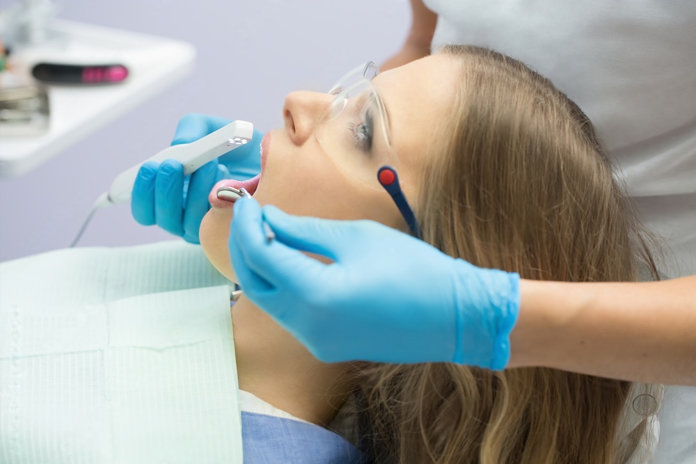
An intraoral camera is a small, pen-shaped device that captures high-resolution photographs and video inside the oral cavity. The camera head typically measures between 5 and 10 millimeters in diameter, allowing it to reach posterior teeth, lingual surfaces, and subgingival margins that are difficult to examine with a mirror alone.
Modern units connect to chairside monitors or laptop screens through USB or wireless protocols. Images appear in real time, giving both the clinician and the patient a clear view of caries, cracks, restorations, soft-tissue lesions, and plaque accumulation.

Key Advances in Intraoral Camera Hardware
Hardware improvements over the last few years have pushed image quality and usability forward significantly. Here are the areas seeing the most progress.
Higher Resolution Sensors
Early intraoral cameras captured images at roughly 640 by 480 pixels. Current models routinely deliver full HD at 1920 by 1080 pixels, and several manufacturers now offer 4K sensors. The jump in resolution means clinicians can zoom into a single cusp tip or margin line and still see fine detail without pixelation.
Autofocus and Depth of Field
Fixed-focus cameras forced operators to hold the wand at a precise distance. Newer autofocus systems lock on within milliseconds, producing sharp images even when the operator moves slightly. Extended depth-of-field algorithms merge multiple focal planes into one composite image, keeping both the buccal wall and the floor of a preparation in focus simultaneously.
LED and Multispectral Lighting
Built-in LED arrays now provide consistent, shadow-free illumination. Some advanced units add near-infrared or fluorescence wavelengths that highlight early enamel demineralization, bacterial biofilm, or cracks invisible under standard white light. This multispectral capability turns the camera from a documentation tool into an active diagnostic instrument.
Software and AI Integration
Hardware alone does not define the value of an intraoral camera. The software that processes and analyzes images is where much of the innovation is happening today.
AI-Assisted Caries Detection
Machine-learning models trained on hundreds of thousands of clinical images can now flag suspected carious lesions, calculus deposits, and defective restorations in real time. The software overlays color-coded indicators on the live feed, drawing the clinician's attention to areas that might otherwise be overlooked during a rapid exam. While final diagnosis remains with the dentist, these AI assistants reduce the risk of missed findings.
Automated Charting and Documentation
Newer platforms link captured images directly to the patient's digital chart. Optical character recognition and tooth-numbering algorithms tag each image to the correct tooth, eliminating manual data entry. Over time, the system builds a visual timeline for every tooth, making it simple to track the progression of a lesion or the condition of a restoration across multiple appointments.
Cloud Storage and Remote Access
Cloud-based image storage allows clinicians to review cases from any location. This is especially useful for group practices, teledentistry consultations, and specialist referrals. A general practitioner can capture images, upload them securely, and have an endodontist or periodontist review the case within hours rather than waiting for an in-person visit.
Teledentistry and Remote Diagnosis
The demand for remote healthcare accelerated rapidly beginning in 2020, and dentistry was no exception. Intraoral cameras are a natural fit for teledentistry because they produce the high-quality visual data that remote clinicians need to evaluate a patient's condition.
In a typical teledentistry workflow, a dental hygienist or assistant captures a standardized set of images using the intraoral camera. Those images are uploaded to a secure platform where a licensed dentist reviews them asynchronously or in a live video session with the patient. The dentist can then recommend treatment, prescribe medication, or schedule an in-office visit if hands-on care is needed.
This model is particularly valuable for underserved communities, rural areas, nursing homes, and school-based dental programs where access to a fully equipped dental office is limited. By placing intraoral cameras in satellite locations, dental organizations can screen far more patients than a single brick-and-mortar clinic could handle.
Improving Patient Education and Case Acceptance
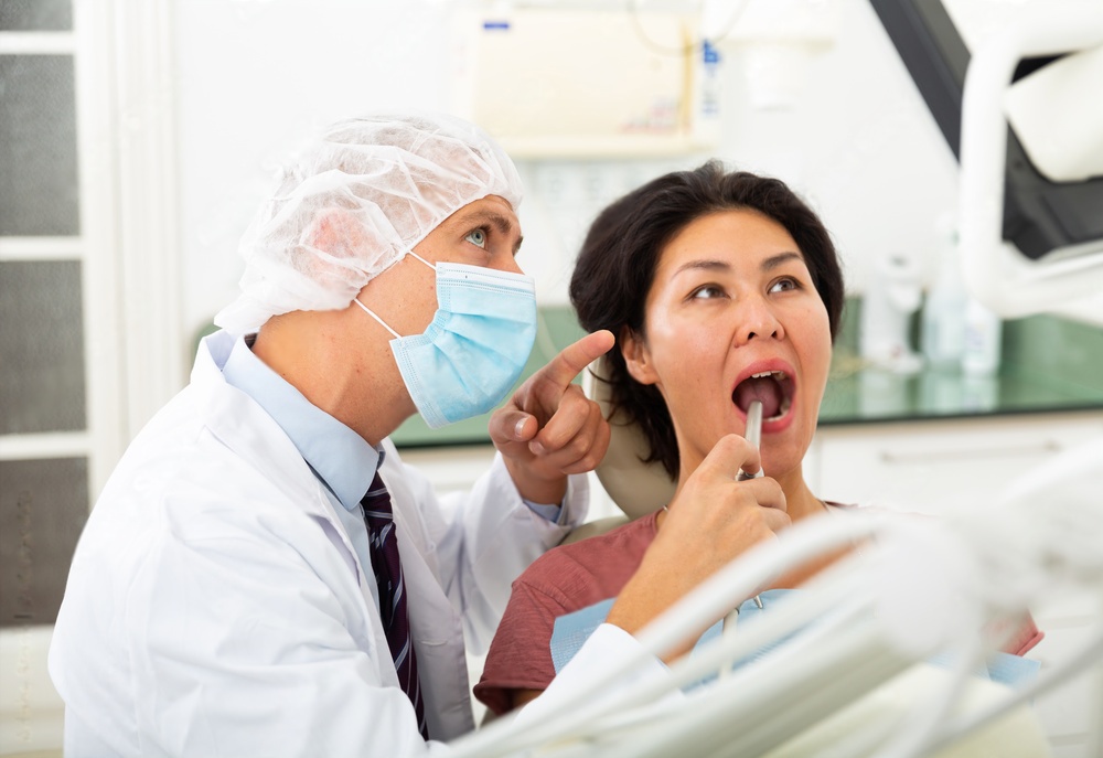
One of the most practical benefits of an intraoral camera has nothing to do with diagnostics. It is about communication. When patients can see a crack running across their molar or a dark shadow beneath an old amalgam, they understand the need for treatment far more quickly than they would from a verbal explanation alone.
Before-and-After Comparisons
Showing a patient a side-by-side image of their tooth before and after a procedure builds trust and satisfaction. It also provides strong documentation in the event of a dispute or insurance inquiry. Many practice management platforms now generate these comparisons automatically.
Treatment Simulation
Emerging software tools overlay predicted outcomes onto the live camera image. A patient considering porcelain veneers, for example, can see a simulated result on screen before committing to the procedure. While still in its early stages, this technology is improving case acceptance rates in cosmetic dentistry.
Choosing an Intraoral Camera for Your Practice
With dozens of models on the market, selecting the right intraoral camera requires evaluating several factors.
- Image resolution: Look for at least full HD. 4K is a worthwhile upgrade if your workflow involves detailed documentation or presentations.
- Connectivity: USB models are simple and reliable. Wireless models offer more freedom of movement but require charging and occasional signal troubleshooting.
- Software compatibility: Confirm that the camera integrates with your existing practice management and imaging software before purchasing.
- Infection control: Disposable sheaths should be readily available and affordable. Autoclavable tips are an alternative but add processing time.
- Ergonomics: A lightweight, well-balanced wand reduces hand fatigue during long sessions. Test several models in hand before making a final decision.
Integrating Cameras with Other Digital Workflows
Intraoral cameras work best when they are part of a connected digital workflow rather than a standalone device. Pairing camera images with cone-beam computed tomography scans, digital impressions, and CAD/CAM milling systems gives clinicians a complete picture of each case. For example, a camera image can confirm the marginal fit of a milled zirconia crown chairside, reducing remakes and saving lab time.
Dental laboratories also benefit from receiving detailed intraoral photographs alongside digital impressions. When a technician can see the shade, texture, and gingival contour in a high-resolution photo, the final restoration is more likely to match the patient's expectations on the first try. Pairing the right abrasive and finishing tools with accurate imaging data leads to predictable, high-quality results.
What to Expect in the Next Five Years
Several trends are likely to shape intraoral camera development through 2030 and beyond.
| Trend | Expected Impact |
|---|---|
| On-device AI processing | Faster analysis without cloud latency; works offline in remote settings |
| Hyperspectral imaging | Detection of early soft-tissue pathology and enamel mineral loss |
| Augmented reality overlays | Real-time guidance during preparations and margin placement |
| Miniaturized sensors | Smaller camera heads for pediatric and limited-opening patients |
| Unified platforms | Single software hub for camera, CBCT, scanner, and CAD/CAM data |
These developments point toward a future where the intraoral camera is not just a documentation device but an active clinical partner that guides decision-making in real time.
Practical Tips for Getting More from Your Current Camera
You do not need to wait for next-generation hardware to improve the value you get from intraoral imaging. A few simple habits can make a noticeable difference.
- Capture a baseline set of images for every new patient. This creates a reference point for all future comparisons.
- Use consistent angulation and lighting for follow-up images so changes are easy to spot.
- Show images to patients during the exam, not just at the end. Real-time viewing increases engagement and trust.
- Store images in a structured folder system tagged by tooth number and date for fast retrieval.
- Review your camera manufacturer's firmware updates regularly. Performance improvements and bug fixes are often released quarterly.
Intraoral cameras have already moved well beyond simple snapshots. As AI, connectivity, and sensor technology continue to mature, these small devices will play an even larger role in how dental professionals diagnose, plan, and communicate treatment. Investing in a quality intraoral camera today, and building workflows around it, positions your practice to take full advantage of the tools that are coming next. For related reading on digital workflows in modern dental practice, see our guide on enhancing diagnosis with dental photography lamps.