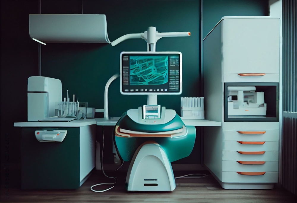
The Digital Revolution in Dental Diagnostics
Dental sensor technology has evolved dramatically over the past two decades, transforming from bulky film-based systems to sleek digital sensors that deliver instant results. Yet we stand at the beginning rather than the end of this revolution. Emerging sensor technologies promise to fundamentally change how we diagnose, plan treatments, and interact with patients. Understanding these developments helps practitioners prepare for the rapidly approaching future of dentistry.
The convergence of artificial intelligence, improved sensor miniaturization, wireless connectivity, and advanced materials science creates unprecedented opportunities. These technologies will not replace clinical judgment but rather enhance our diagnostic capabilities and simplify workflows in ways previously unimaginable.
Artificial Intelligence and Machine Learning Integration
AI-powered diagnostic systems represent perhaps the most transformative application of sensor technology. Current dental sensors capture images, but the interpretation remains largely manual. Next-generation systems will analyze sensor data in real-time, identifying potential issues and suggesting treatment approaches based on massive datasets of previous cases.
How AI Enhances Sensor Diagnostics
- Automated detection of early caries before they're visible to the human eye
- Identification of bone density changes indicating periodontal disease progression
- Analysis of radiographic patterns suggesting pathology
- Comparison of current images with patient history to track changes over time
- Prediction of treatment outcomes based on anatomical measurements
- Reduction of false positives through multi-factor analysis
- Standardization of diagnostic criteria across practitioners
These AI systems will function as diagnostic assistants rather than replacements for clinical judgment. A sensor might flag a suspicious area and provide a confidence level, prompting closer examination. Over time, the system learns from confirmed diagnoses, continuously improving accuracy. Early implementations already show promise in detecting interproximal caries and periapical lesions with accuracy matching or exceeding experienced practitioners.
The implications extend beyond individual patient care. Aggregated sensor data from thousands of practices could identify population-level trends, helping public health officials target preventive programs more effectively. Insurance companies may use sensor-verified diagnostics to simplify claims processing, reducing administrative burden on practices.
Advanced 3D Imaging Sensors
While CBCT technology provides three-dimensional views, it requires dedicated equipment, significant radiation exposure, and substantial cost. Emerging 3D sensor technologies aim to deliver volumetric data with lower radiation, smaller footprints, and integration into routine workflows.
Next-Generation 3D Sensor Capabilities
| Technology | Application | Advantage |
|---|---|---|
| Photon-counting sensors | Low-dose 3D imaging | 80% radiation reduction versus current CBCT |
| Multi-angle synthesis | 3D reconstruction from 2D images | No additional equipment needed |
| Optical coherence tomography | Soft tissue imaging | No ionizing radiation |
| Ultrasound sensors | Periodontal pocket mapping | Real-time 3D visualization |
| Structured light scanning | Preparation margin detection | Immediate digital impressions |
These technologies will enable practitioners to visualize anatomy in three dimensions routinely rather than only for complex cases. Imagine reviewing a standard checkup radiograph and instantly rotating the view to examine interproximal areas from multiple angles, or evaluating root canal anatomy in three dimensions before beginning treatment. This level of detail improves treatment planning while reducing procedural complications.
The integration with CAD/CAM workflows becomes seamless when sensors natively output 3D data. Treatment planning, virtual wax-ups, and milling instructions flow from the same sensor data captured chairside, eliminating transfer errors and improving precision.
Wireless and Battery-Free Sensor Systems
Current digital sensors require cables that limit positioning flexibility and create infection control challenges. Wireless sensors already exist, but battery requirements add bulk and create reliability concerns. Emerging technologies eliminate these limitations entirely.
Wireless Technology Advances
Next-generation sensors use energy harvesting to power themselves from the x-ray exposure itself, eliminating batteries completely. Others employ ultra-low-power wireless protocols that enable years of operation from tiny integrated batteries. Some designs use inductive charging integrated into sensor holders, ensuring the sensor is always ready without manual intervention.
The clinical advantages extend beyond convenience. Thinner sensors without battery compartments improve patient comfort, particularly for children and patients with small mouths. Wireless operation eliminates cable strain, extending sensor lifespan and reducing replacement costs. Infection control improves when barriers don't need to accommodate cables, and positioning becomes more flexible when cable routing doesn't constrain sensor placement.
Wireless sensors also enable new imaging protocols. Multiple sensors could be positioned simultaneously, capturing several views with a single exposure series. This approach reduces total radiation while improving diagnostic information, particularly for evaluating TMJ anatomy or full-arch implant planning.
Internet of Things Integration
IoT technology connects devices in ways that create value beyond their individual capabilities. Dental sensors are beginning to participate in this ecosystem, communicating not just with imaging software but with the entire practice management infrastructure.
IoT-Enabled Sensor Applications
- Automatic patient identification when sensors are positioned, eliminating mislabeled images
- Integration with electronic health records for instant image filing and retrieval
- Predictive maintenance alerts based on sensor usage patterns and performance metrics
- Automatic exposure parameter optimization based on patient size and diagnostic needs
- Quality assurance monitoring flagging suboptimal images before the patient leaves
- Inventory tracking of sensors, barriers, and supplies with automatic reordering
- Remote diagnostics enabling manufacturer support without shipping sensors for service
Consider a typical workflow in an IoT-enabled practice. The sensor recognizes it's in operatory three based on local beacons. It identifies the patient from the appointment schedule. It recommends exposure settings based on the patient's age, size, and diagnostic history. After exposure, it analyzes image quality and suggests retakes if needed. The finalized images automatically appear in the patient record with proper labeling and dating. Usage data flows to the practice management system for billing and to the manufacturer for warranty tracking.
This level of integration reduces errors, simplifies workflows, and frees staff time for patient interaction rather than data entry. The technology handles routine tasks while practitioners focus on clinical decision-making.
Real-Time Caries Detection Sensors
Current caries detection relies primarily on visual examination, tactile exploration, and radiographic interpretation. Emerging sensor technologies detect caries through multiple physical mechanisms, identifying demineralization before structural changes become visible.
Fluorescence-based sensors illuminate teeth with specific wavelengths and analyze the reflected light patterns. Carious lesions fluoresce differently than healthy enamel, enabling detection of early demineralization. Electrical impedance sensors measure how electric current flows through tooth structure, with carious areas showing different conductivity than sound enamel. Optical coherence tomography uses near-infrared light to create cross-sectional images of tooth structure, revealing subsurface lesions invisible to conventional radiography.
Integration with Treatment Decisions
These sensors don't just detect caries but quantify severity, enabling more nuanced treatment decisions. A lesion scoring 2 on a 10-point scale might warrant fluoride treatment and monitoring, while a score of 8 clearly requires restoration. This objectivity reduces both overtreatment of arrested lesions and undertreatment of active decay.
When combined with AI analysis of risk factors, these sensors enable personalized caries prevention programs. A patient showing early demineralization in specific areas receives targeted interventions rather than generic recommendations. Follow-up scans track whether prevention strategies are succeeding, allowing real-time protocol adjustments.
For patients, real-time caries detection sensors provide compelling visual evidence. Seeing a color-coded map of tooth demineralization makes abstract concepts concrete, improving treatment acceptance and motivating better home care. This technology transforms patient education from telling to showing.
Enhanced Image Resolution and Clarity
While current digital sensors provide adequate resolution for most applications, emerging technologies push boundaries significantly higher. Ultra-high-resolution sensors capture details impossible to visualize previously, enabling earlier detection of subtle pathology.
| Resolution Class | Line Pairs per mm | Primary Application |
|---|---|---|
| Current standard sensors | 16-20 lp/mm | General diagnostic imaging |
| High-resolution sensors | 25-30 lp/mm | Endodontic anatomy |
| Ultra-high-resolution sensors | 35-45 lp/mm | Research and specialized diagnostics |
| Emerging nanosensors | 50+ lp/mm | Microscopic defect detection |
These resolution improvements enable visualization of root canal anatomy details that guide treatment decisions. Isthmuses, lateral canals, and accessory foramina become clearly visible, reducing surprises during instrumentation. Margin quality assessment for indirect restorations improves when sensors can resolve 10-micron gaps rather than 50-micron minimums.
The challenge lies in data management. Ultra-high-resolution images create massive files requiring substantial storage and processing power. Cloud-based systems and improved compression algorithms address these concerns, making ultra-high-resolution imaging practical for routine use rather than limiting it to research applications.
Integration with CAD/CAM Workflows
The boundary between diagnostic sensors and design tools continues to blur. Sensors that capture both radiographic data and surface geometry enable seamless transitions from diagnosis to treatment planning to restoration fabrication.
Unified Digital Workflow
Imagine a complete sensor system that captures a periapical radiograph, maps the preparation margin in three dimensions, records occlusal relationships, and determines tooth shade - all from a single device. This data feeds directly into CAD software where the restoration design considers not just the visible preparation but the underlying anatomy visible radiographically. The milling unit receives instructions optimized for the specific material based on measured properties, and the appropriate milling burs are automatically selected.
This integration eliminates data transfer errors that occur when moving between systems. Preparation dimensions measured by the sensor match exactly what the CAD software receives. Color values captured at acquisition remain consistent through design and fabrication. The result is restorations that fit better, match more accurately, and require fewer adjustments.
For practices already invested in digital workflows, sensor integration represents the final piece connecting diagnosis to treatment to fabrication. For those still using traditional methods, these unified systems provide a compelling reason to transition, delivering return on investment through reduced remake rates and improved efficiency. Learn more about the intersection of technology and restoration in our article on the latest in dental sensor tech.
Multi-Spectral and Hyperspectral Imaging
Conventional sensors capture images at specific x-ray energies, essentially seeing in one color. Multi-spectral sensors capture images at multiple energies simultaneously, while hyperspectral sensors record dozens or hundreds of energy bands. This additional data enables material differentiation impossible with single-energy imaging.
The practices that thrive will be those that thoughtfully adopt technologies aligned with their clinical and business goals. Success requires balancing innovation with practicality, investing in capabilities that improve patient care and practice efficiency while avoiding technology for its own sake. For additional insights into maintaining your imaging equipment, see our guide on improving dental x-ray image quality with sensors.
Ultimately, dental sensor technology serves one purpose: enabling practitioners to provide better care for patients. The most successful implementations will be those that keep this goal central, using technology as a tool to enhance rather than replace the clinical judgment, technical skill, and patient relationships that define excellent dentistry. The future of dental sensors is not about replacing the practitioner but about providing better information to support clinical decisions and improve patient outcomes.
As these technologies mature and become more accessible, every practice will need to determine how to integrate them into existing workflows. Those decisions will shape the patient experience, clinical outcomes, and practice success for years to come. Understanding the possibilities helps practitioners make informed choices that position their practices for long-term success in an increasingly digital profession.