Understanding When Dental Implants Need Replacement
Dental implants have transformed restorative dentistry with success rates exceeding 95% over ten years. However, certain conditions require implant component replacement or complete implant removal. Recognizing these signs early prevents complications and preserves bone structure for future treatment options.
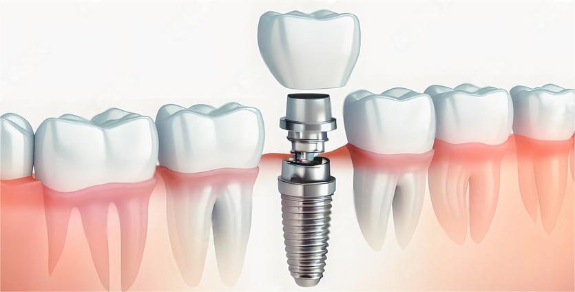
Modern implant systems consist of three primary components: the titanium fixture embedded in bone, the abutment connecting to the fixture, and the prosthetic crown or bridge. Each component has distinct failure patterns requiring different intervention approaches.
Critical Signs Your Dental Implant Requires Replacement
Peri-Implantitis and Infection
Peri-implantitis represents the leading cause of late implant failure. This inflammatory condition affects tissues surrounding the implant, causing progressive bone loss. Clinical indicators include bleeding on probing, suppuration, increased probing depths exceeding 5mm, and radiographic bone loss beyond physiological remodeling.
Early peri-implant mucositis affects only soft tissues and responds to non-surgical treatment. Advanced peri-implantitis with bone loss requires surgical intervention and potentially implant removal if decontamination procedures fail to arrest disease progression.
Prosthetic Component Wear and Fracture
Crown and bridge materials deteriorate over 10-15 years of functional loading. Porcelain fractures, excessive occlusal wear, margin discoloration, and loss of anatomical contour indicate prosthetic replacement needs. These issues compromise function and aesthetics without necessarily affecting the underlying implant fixture.
Metal framework fatigue rarely occurs with quality materials but becomes problematic in thin cross-sections or excessive cantilever designs. Radiographic examination reveals framework fractures before complete separation occurs.
Abutment Loosening and Screw Complications
Abutment screw loosening creates a characteristic clicking sensation during function and increases bacterial infiltration risk. Repeated loosening episodes indicate improper torque values, damaged screw threads, or occlusal overload requiring correction.
Screw fracture within the implant body presents significant retrieval challenges. Specialized removal kits with reverse-threaded instruments extract fractured components, but success depends on fracture location and remaining thread engagement.
Progressive Bone Loss Patterns
Physiological bone remodeling averages 1-1.5mm during the first year, then less than 0.2mm annually. Bone loss exceeding these parameters suggests biomechanical overload, infection, or systemic factors affecting osseointegration maintenance.
Vertical bone defects create deep pockets harboring bacteria. Horizontal bone loss reduces implant support circumference. Both patterns compromise long-term stability and may necessitate implant removal with bone grafting before replacement fixture placement.
Diagnostic Procedures for Implant Assessment
| Diagnostic Method | Information Provided | Frequency |
|---|---|---|
| Periapical Radiographs | Bone levels, fixture integrity, component seating | Annually or as indicated |
| CBCT Imaging | 3D bone architecture, fenestrations, dehiscences | Pre-surgical planning, complex cases |
| Clinical Probing | Soft tissue health, pocket depths, bleeding | Each maintenance visit |
| Percussion Testing | Osseointegration status, mobility detection | Symptomatic evaluation |
| Occlusal Analysis | Force distribution, premature contacts | Initial placement and annually |
complete documentation establishes baseline parameters for monitoring changes over time. Digital imaging allows precise measurement comparisons between appointments, identifying subtle progressive changes requiring intervention.
Mobility and Resonance Frequency Analysis
Clinical mobility indicates complete osseointegration loss requiring implant removal. Even slight movement under lateral force suggests failed integration. Electronic devices measuring resonance frequency quantify implant stability objectively, detecting early integration loss before clinical mobility appears.
Replacement Options and Treatment Approaches
Component-Level Replacement
Prosthetic replacement without fixture disturbance represents the simplest intervention. Crown and bridge removal, abutment replacement, and new prosthetic fabrication require 2-3 appointments over several weeks. This approach preserves the osseointegrated fixture while addressing aesthetic and functional concerns.
Abutment replacement corrects angulation issues, changes prosthetic emergence profiles, and eliminates damaged connection components. Modern abutment designs offer custom tissue contouring and improved material options like zirconia for anterior aesthetics.
Complete Implant Removal and Replacement
Failed fixture removal requires careful technique preserving surrounding bone. Trephine burs create a circumferential osteotomy separating the implant from bone without excessive trauma. Counter-rotation with removal instruments extracts the fixture maintaining bone architecture.
Immediate replacement in fresh extraction sites succeeds when adequate bone remains and infection is controlled. This approach reduces treatment time but requires precise case selection based on bone quality, infection status, and anatomical position.
Staged Replacement with Bone Regeneration
Significant bone loss necessitates grafting procedures before implant placement. Autogenous bone, allografts, xenografts, or synthetic materials restore ridge dimensions over 4-6 months healing. Guided bone regeneration with barrier membranes maintains graft volume during healing.
Sinus augmentation addresses maxillary posterior bone deficiency. Ridge splitting techniques expand narrow ridges horizontally. Block grafts restore substantial defects. Each technique requires specific healing periods before implant placement.
Treatment Timeline Expectations
Understanding realistic timeframes helps patients plan appropriately for implant replacement procedures:
- Prosthetic-Only Replacement: 2-4 weeks for crown fabrication and delivery after impressions
- Abutment and Prosthetic Replacement: 3-5 weeks including healing time after abutment placement
- Immediate Fixture Replacement: 3-6 months for osseointegration plus 4-8 weeks for prosthetic fabrication
- Staged Replacement with Grafting: 4-6 months graft healing, 3-6 months implant integration, 4-8 weeks prosthetic completion
- Complex Bone Reconstruction: 6-12 months total treatment time depending on graft requirements and healing response
Individual healing capacity, medical conditions, and smoking status influence these timeframes. Diabetic patients and smokers typically require extended healing periods for optimal outcomes.
Preventing Implant Failure and Component Wear
Maintenance Protocol Essentials
Professional maintenance every 3-4 months prevents peri-implant disease through mechanical debridement and infection monitoring. Plastic or titanium instruments avoid implant surface damage during cleaning. Antimicrobial irrigation reduces bacterial colonization in peri-implant pockets.
Home care requires specific techniques addressing implant anatomy. Interproximal brushes access embrasure spaces beneath prosthetic pontics. Water flossers with specialized tips irrigate sulcular areas. Low-abrasive toothpastes prevent prosthetic surface scratching that promotes plaque retention.
Occlusal Management and Night Guards
Occlusal equilibration eliminates premature contacts creating excessive force on implant components. Proper contacts in centric relation and excursive movements distribute forces physiologically. Implants lack periodontal ligament proprioception, making them vulnerable to force concentration without natural dampening.
Bruxism and clenching habits dramatically increase implant complications. Custom night guards protect prosthetic materials and reduce biomechanical stress on fixtures and bone. Hard acrylic guards offer superior protection compared to soft materials that may increase grinding activity.
Systemic Health Optimization
Diabetes control with HbA1c below 7% significantly improves implant success rates. Hyperglycemia impairs wound healing and infection resistance. Medication management including bisphosphonates requires coordination with physicians regarding timing and alternative therapies.
Smoking cessation remains the single most important modifiable risk factor. Nicotine reduces blood flow impairing osseointegration and maintenance. Patients who quit smoking before implant procedures demonstrate markedly improved outcomes compared to continuing smokers.
Post-Replacement Care and Long-Term Success
Initial healing following implant replacement requires careful protection. Soft diet for 6-8 weeks prevents excessive loading during osseointegration. Chlorhexidine rinses twice daily control bacterial levels during soft tissue healing. Suture removal at 10-14 days allows tissue inspection and secondary closure if indicated.
Prosthetic delivery includes occlusal verification with shimstock and articulating paper in multiple jaw positions. Radiographic confirmation ensures complete component seating. Patient education covering cleaning techniques and maintenance schedules establishes long-term success foundations.
Annual radiographic evaluation monitors bone levels around replacement implants. Stability testing confirms ongoing osseointegration. Early intervention for emerging problems prevents catastrophic failures requiring extensive reconstruction.
Working with Your Dental Team
Complex implant replacement cases benefit from specialist coordination. Periodontists manage peri-implant disease and bone grafting. Oral surgeons perform difficult extractions and ridge reconstruction. Prosthodontists design optimal prosthetic solutions for challenging situations.
Communication between specialists ensures cohesive treatment planning. Digital files sharing implant positions, bone architecture, and prosthetic designs simplifies the process. Patient involvement in decision-making regarding timing, materials, and cost considerations creates realistic expectations.
When implant complications arise, prompt professional evaluation determines appropriate intervention. Early treatment preserves bone and soft tissue, maintaining options for successful replacement. Delaying necessary treatment often complicates eventual solutions and compromises final outcomes.
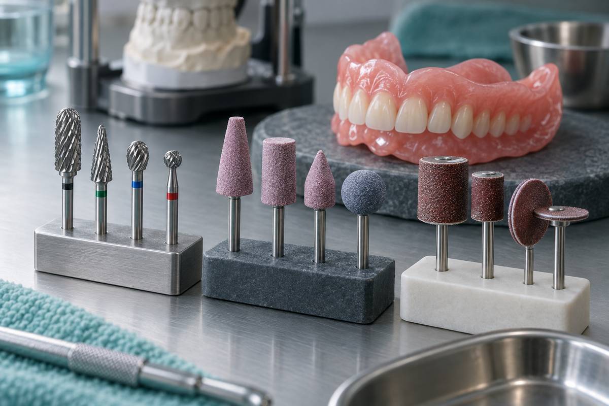
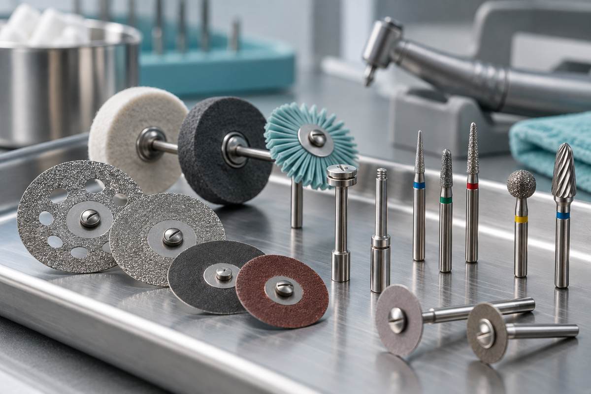
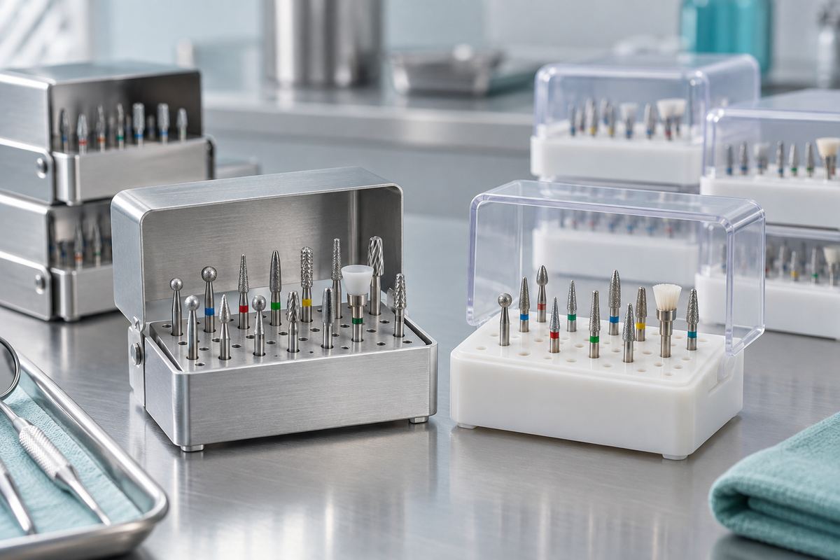
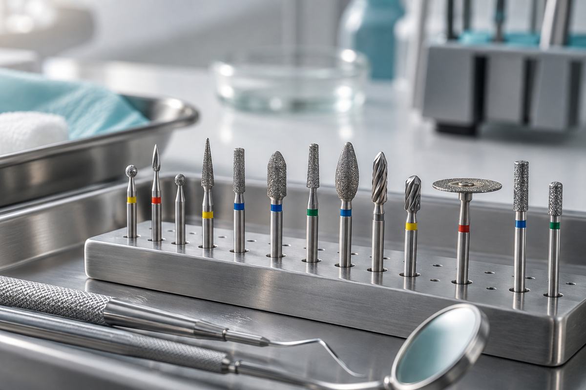
For preparing implant sites and managing bone during replacement procedures, specialized surgical burs provide precision and control. Additionally, proper finishing and contouring of provisional and final restorations requires quality tungsten carbide burs for optimal results.
Learn more about maintaining dental implants through our guide on the latest trends in dental implant technology and explore how technology is revolutionizing tooth replacement for advanced treatment options.