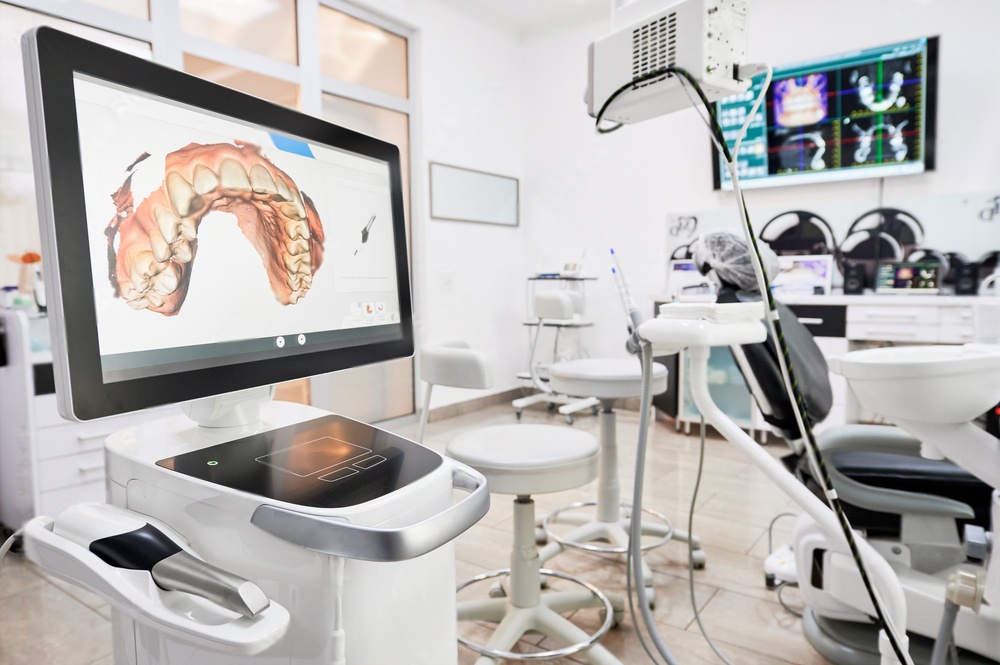
How Dental Sensor Technology Is Transforming Clinical Diagnostics
Dental imaging has undergone a fundamental shift over the past two decades. Film-based radiography, once the only option for visualizing tooth structures, has given way to digital sensors that produce higher-quality images at lower radiation doses. The pace of innovation continues to accelerate, with new sensor designs offering capabilities that were not commercially viable even five years ago.
This article examines the current state of dental sensor technology, covering the major categories of digital imaging sensors, their clinical applications, and the practical considerations that guide purchasing decisions for dental practices.
Digital Radiography Sensors: The Foundation of Modern Dental Imaging
Digital radiography sensors replaced traditional film by converting X-ray energy directly into electronic signals. Two primary technologies dominate this space:
- CCD (Charge-Coupled Device) sensors use a silicon chip to capture X-ray photons and convert them to an electrical charge. They produce high-resolution images with excellent contrast.
- CMOS (Complementary Metal-Oxide Semiconductor) sensors offer lower power consumption and faster image readout. Manufacturing costs are lower, which has made CMOS the more common choice in newer sensor designs.
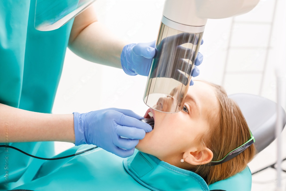
Both technologies deliver immediate image display on a chairside monitor, eliminating the processing time associated with film. More importantly, digital sensors require 50 to 80 percent less radiation than conventional film to produce a diagnostic image.
Sensor Size and Patient Comfort
Early digital sensors were noticeably thicker than film packets, causing patient discomfort during placement. Current-generation sensors have addressed this limitation significantly. Active pixel areas now measure as thin as 5.5 mm in some models, approaching the profile of traditional film. Rounded corners and flexible cable attachments further improve patient tolerance.
Sensor sizing follows the standard film classification:
| Size | Designation | Primary Use |
|---|---|---|
| Size 0 | Pediatric | Primary dentition and small adult mouths |
| Size 1 | Anterior | Anterior periapical views in adults |
| Size 2 | Standard | Posterior periapical and bitewing views |
Practices that serve a mixed patient population typically invest in at least two sensor sizes to ensure proper coverage across all areas of the mouth.
Intraoral Cameras: Visual Documentation at Chairside
Intraoral cameras serve a different purpose than radiographic sensors. Rather than capturing X-ray images, they photograph visible tooth surfaces, soft tissue, and restorations in high definition. Their clinical value extends across several areas:
- Patient communication becomes more effective when patients can see exactly what the clinician sees. A magnified image of a cracked filling or inflamed gingiva is far more persuasive than a verbal description alone.
- Documentation for insurance claims and legal records benefits from timestamped photographic evidence of existing conditions.
- Treatment monitoring allows clinicians to compare images over time, tracking the progression or resolution of lesions, restorations, and soft tissue conditions.
Current wireless intraoral cameras weigh under 50 grams and connect via WiFi or Bluetooth to practice management software. Some models include autofocus and LED illumination that adjusts automatically based on the distance to the target surface.
Integration With Practice Management Systems
The clinical value of an intraoral camera depends heavily on how well it integrates with existing software. Leading camera systems support TWAIN and DirectShow protocols, allowing captured images to flow directly into the patient record without manual file transfer. DICOM compatibility ensures that images from different devices can be stored and retrieved in a standardized format.
CBCT: Three-Dimensional Imaging for Complex Cases
Cone Beam Computed Tomography represents the most significant advance in dental imaging technology of the past fifteen years. Unlike conventional panoramic radiographs that produce a two-dimensional projection, CBCT captures a volumetric dataset that can be viewed in axial, coronal, and sagittal planes.
CBCT has become the standard of care for several clinical scenarios:
- Implant planning requires precise measurement of bone height, width, and density at the proposed implant site. CBCT data integrates with surgical planning software to generate drill guides.
- Endodontic assessment benefits from the ability to visualize root canal morphology, periapical lesions, and root fractures that periapical radiographs may miss.
- Impacted teeth evaluation provides three-dimensional localization of impacted third molars relative to the inferior alveolar nerve canal.
- Orthodontic analysis enables accurate cephalometric measurements and assessment of airway dimensions.
- TMJ evaluation reveals bony changes in the condyle and fossa that are not visible on standard radiographs.
Field of view (FOV) selection is critical in CBCT imaging. Smaller FOV scans (5x5 cm) deliver higher resolution with lower radiation for focused diagnostic tasks, while larger FOV scans (16x13 cm) capture the full craniofacial complex when comprehensive data is needed.
Fluorescence and Transillumination: Early Decay Detection
Beyond radiographic imaging, optical sensor technologies have opened new pathways for detecting caries at their earliest stages.
Quantitative Light-Induced Fluorescence (QLF)
QLF devices project a specific wavelength of light onto the tooth surface. Healthy enamel fluoresces at a predictable intensity, while demineralized areas show reduced fluorescence. This allows clinicians to detect white spot lesions and incipient caries before they become visible on radiographs. The technology is particularly valuable for monitoring remineralization therapy and assessing the effectiveness of preventive interventions.
Near-Infrared Transillumination (NIRT)
NIRT passes near-infrared light through tooth structure. Carious lesions and cracks scatter the light differently than intact enamel, creating visible contrast in the captured image. Unlike radiography, NIRT involves zero ionizing radiation, making it suitable for frequent monitoring without dose concerns. It has proven especially effective for detecting interproximal caries in posterior teeth.
Laser Fluorescence (DIAGNOdent)
Laser fluorescence devices measure the fluorescence emitted by bacterial metabolites within carious lesions. A numeric readout provides an objective measurement that supplements clinical and radiographic findings. While not a replacement for radiographs, laser fluorescence adds a quantitative dimension to caries diagnosis that reduces reliance on subjective visual assessment.
Choosing the Right Sensor for Your Practice
Selecting imaging equipment involves balancing diagnostic capability, workflow efficiency, and budget. The following factors should guide the evaluation process:
- Image resolution measured in line pairs per millimeter (lp/mm). Diagnostic radiography requires a minimum of 20 lp/mm; most current sensors exceed this threshold.
- Sensor durability including bite force resistance and cable longevity. Replacement costs for damaged sensors can be substantial.
- Software compatibility with your existing practice management system. Proprietary image formats create vendor lock-in that complicates future equipment changes.
- Warranty and support terms, including turnaround time for sensor replacement under warranty.
- Total cost of ownership accounting for the sensor, software license, support contract, and replacement units over a five-year period.
Dental practices that invest in quality diagnostic equipment also benefit from using precision instruments during treatment. Diamond dental burs complement advanced imaging by enabling the accurate cavity preparations that digital diagnostics make possible.
Infection Control Considerations for Digital Sensors
Digital sensors cannot be autoclaved. Infection control protocols rely on barrier sleeves that cover the sensor and cable during patient contact. After each use, the barrier is removed and the sensor is wiped with an intermediate-level disinfectant.
Key infection control practices include:
- Using FDA-cleared barrier sleeves that fit the specific sensor model
- Inspecting barriers for tears before and after each use
- Disinfecting the sensor with manufacturer-approved solutions only
- Storing sensors in a clean, dry location between uses
- Documenting barrier failures and implementing corrective action
Failure to follow proper infection control protocols can result in cross-contamination and sensor damage from exposure to incompatible chemicals.
The Future of Dental Sensor Technology
Several emerging developments suggest where dental sensor technology is headed in the near term. Artificial intelligence algorithms are being trained to detect caries, periapical pathology, and bone loss from radiographic images with accuracy rates comparable to experienced clinicians. Photon-counting detector technology promises to further reduce radiation dose while improving image contrast. Wireless sensor designs that eliminate cable failures are moving from prototype to production.
For a deeper look at where sensor innovation is heading, see our article on future applications for dental sensor technology. Additional guidance on sensor selection can be found in our guide to finding your perfect dental X-ray sensor match.
Summary
Digital sensor technology has made dental imaging faster, safer, and more informative. From intraoral radiographic sensors that reduce radiation exposure to CBCT systems that reveal three-dimensional anatomy, today's imaging tools give clinicians diagnostic information that was inaccessible a generation ago. Optical detection methods add another layer of capability, catching decay at stages where conservative intervention can preserve tooth structure. As these technologies continue to improve, the standard of diagnostic care in dentistry will advance with them.