Latest Dental Implant Technology Trends You Should Know
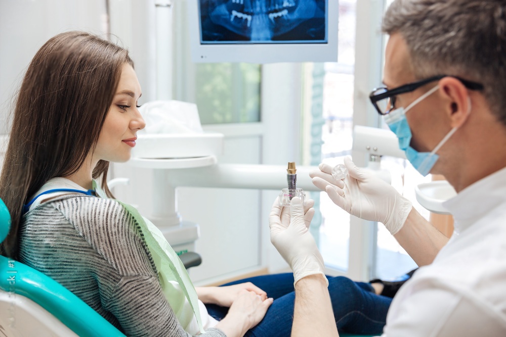
Dental implant procedures have changed dramatically in recent years. New materials, digital workflows, and robotic-assisted surgery now allow clinicians to place implants with greater accuracy and shorter healing times. For patients, these advances mean less discomfort, faster recovery, and restorations that look and feel like natural teeth.
This guide covers the most significant dental implant technology trends shaping modern practice, from advanced titanium alloys to computer-guided robotic placement.
Advanced Titanium Alloys for Stronger Implants
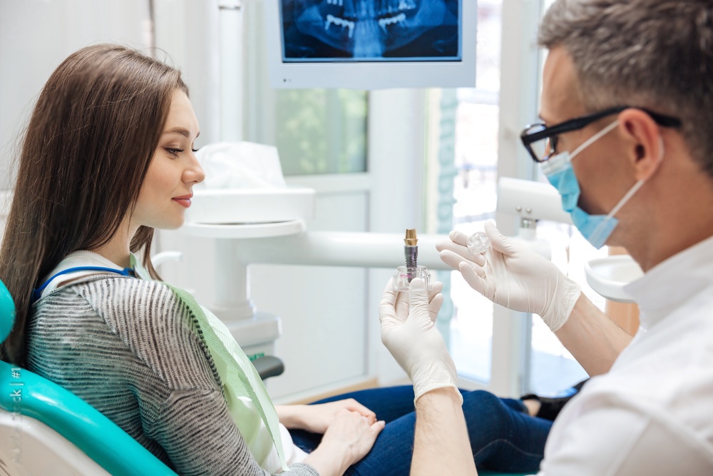
Titanium has been the standard implant material for decades, but recent metallurgical developments have produced alloys that outperform conventional commercially pure (cp) titanium in several ways.
Grade 5 Titanium (Ti-6Al-4V)
Grade 5 titanium alloy combines titanium with aluminum and vanadium to produce a material that is significantly stronger than cp titanium. This increased strength allows manufacturers to design narrower-diameter implants without sacrificing structural integrity. Narrower implants preserve more of the surrounding alveolar bone, which is particularly beneficial for patients with limited bone volume.
Roxolid and Next-Generation Alloys
Straumann's Roxolid alloy, a titanium-zirconium blend, offers roughly 50% greater fatigue strength compared to cp Grade 4 titanium. Other manufacturers have introduced similar high-performance alloys that maintain excellent biocompatibility while enabling smaller implant dimensions. These materials are especially useful in the anterior zone, where bone width is often limited and aesthetics are paramount.
Conic (Morse Taper) Connections
Traditional external and internal hex connections have served the industry well, but conic or Morse taper connections represent a meaningful improvement in implant-abutment stability.
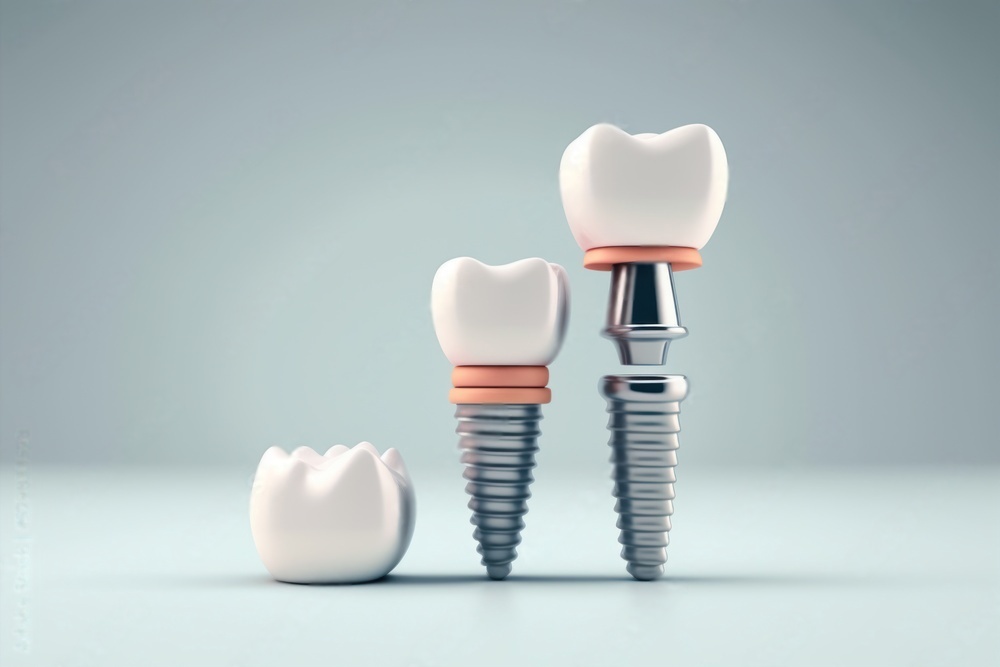
A conic connection uses a precise taper angle, typically between 1.5 and 5 degrees, to create a cold weld between the implant body and the abutment. This friction-fit design offers several clinical advantages:
- Reduced microgap: The tight seal between components minimizes bacterial infiltration at the implant-abutment interface.
- Platform switching effect: The connection geometry shifts stress away from the crestal bone, helping to preserve marginal bone levels.
- Improved stability: The self-locking taper resists loosening even under high occlusal loads.
- Simplified prosthetics: Many conic systems allow for one-position placement, reducing the chance of misalignment during restoration.
Clinical studies have demonstrated that conic connections maintain stable crestal bone levels over five-year follow-up periods, with marginal bone loss averaging less than 0.5 mm.
Laser-Etched and Micro-Textured Surfaces
Surface topography plays a direct role in how quickly and securely an implant integrates with surrounding bone. Modern surface treatment technologies have moved well beyond the simple acid-etched or sandblasted surfaces of earlier generations.
How Laser Etching Works
Laser surface modification uses precisely controlled energy pulses to create micro- and nano-scale features on the implant surface. These features, which include grooves, pits, and pillars at the cellular scale, provide increased surface area for bone cell attachment. The result is faster osseointegration and stronger bone-to-implant contact.
Clinical Benefits of Advanced Surfaces
Research published in the Journal of Dental Research has shown that laser-modified surfaces can reduce initial healing time from the traditional 12 weeks down to 6-8 weeks in many cases. Some manufacturers now combine laser texturing with hydrophilic surface chemistry, which allows blood to wet the implant surface immediately upon placement. This accelerates the initial stages of the healing cascade and can be particularly helpful when treating patients with compromised bone quality.
3D-Printed Custom Implants
Additive manufacturing has moved from a research curiosity to a practical clinical tool. While most standard implants are still machined from titanium bar stock, 3D printing is opening new possibilities for complex cases.
Patient-Specific Implant Design
Using CBCT scan data, engineers can design implants that match a patient's unique anatomy. This is especially valuable for patients with significant bone defects or unusual anatomical features where standard implant sizes may not fit well. The digital file is sent directly to a selective laser melting (SLM) printer that builds the implant layer by layer from titanium powder.
Lattice Structures for Better Integration
One advantage unique to 3D printing is the ability to create internal lattice or scaffold structures within the implant body. These porous architectures mimic the trabecular structure of natural bone and allow bone to grow into the implant rather than simply around it. Early clinical data suggests that lattice-structured implants may achieve higher removal torque values, indicating stronger mechanical integration.
Surgical Guides and Planning
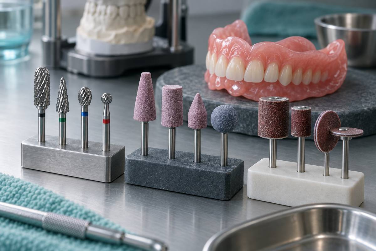
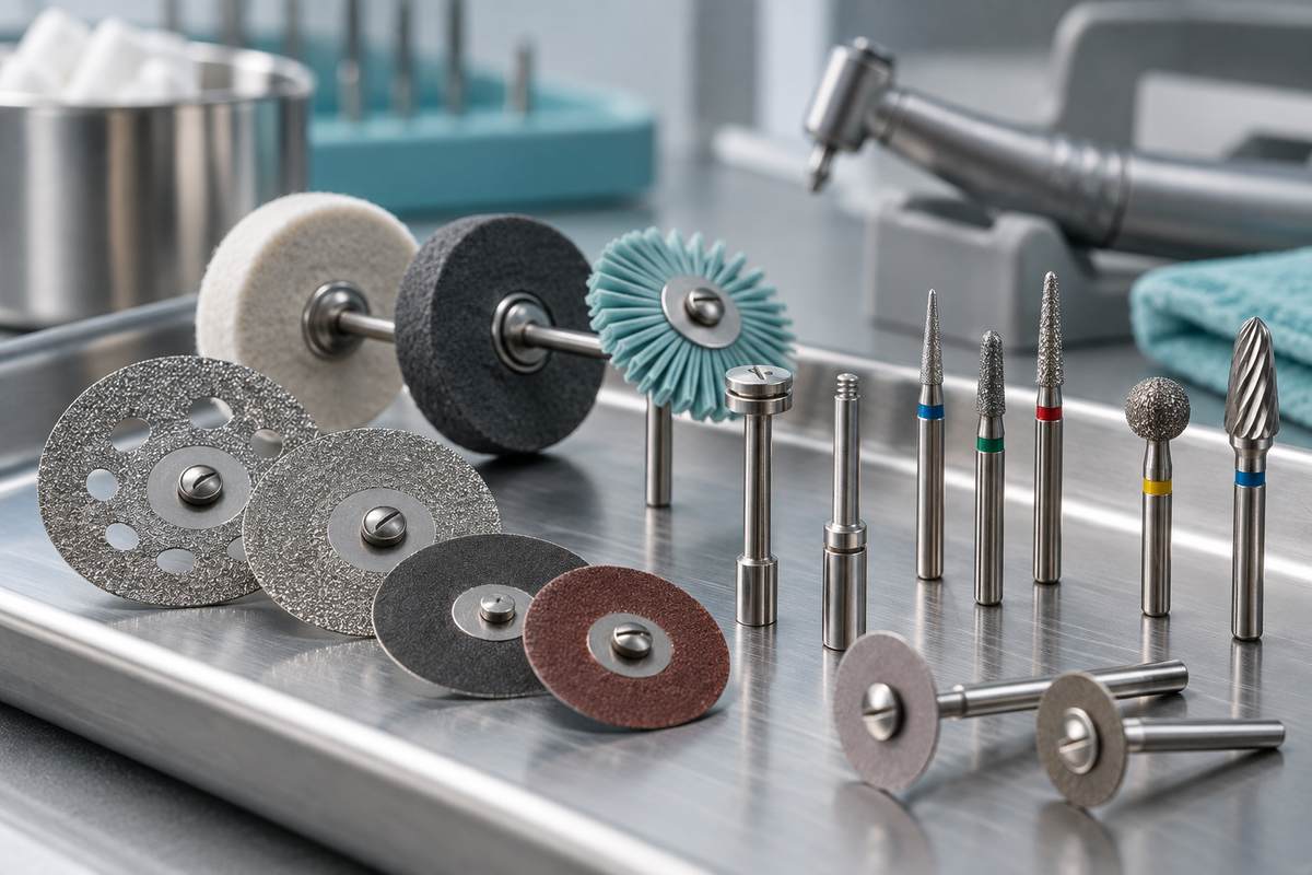
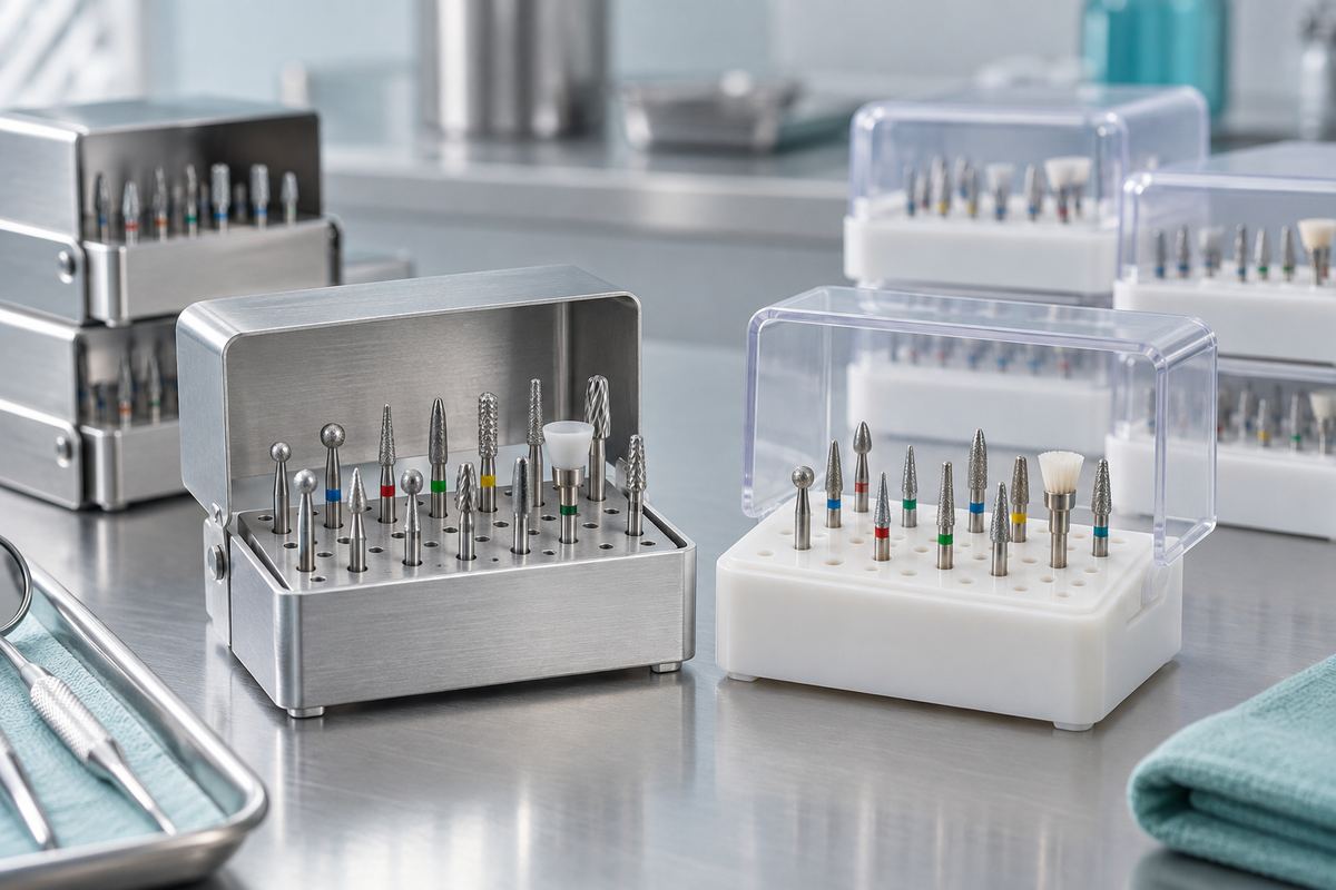
Beyond the implants themselves, 3D printing has become standard for producing surgical drill guides. These guides, printed from biocompatible resin based on virtual implant planning software, ensure that the osteotomy is prepared at the exact angle, depth, and position planned digitally. This reduces surgical time and improves predictability, especially for full-arch cases. Precision placement also depends on high-quality surgical burs that cut cleanly at the planned angles.
Robotic-Assisted Implant Surgery
Robotic systems represent the next step beyond static surgical guides. Several systems are now commercially available and gaining traction in private practice settings.
How Robotic Implant Placement Works
Systems like Yomi (Neocis) and others use a robotic arm that works in conjunction with a pre-operative digital plan. The dentist maintains full control of the handpiece, but the robot provides real-time haptic feedback and physical boundaries that prevent deviation from the planned trajectory. If the surgeon begins to drift off-course, the system applies gentle resistance to redirect the drill path.
Accuracy and Outcomes
A 2024 systematic review in Clinical Oral Implants Research found that robotic-assisted implant placement achieved mean angular deviation of 1.2 degrees and mean apical deviation of 0.4 mm, both significantly better than freehand placement. This level of precision is particularly important when placing implants near critical anatomical structures such as the inferior alveolar nerve or the maxillary sinus floor.
| Placement Method | Angular Deviation | Apical Deviation |
|---|---|---|
| Freehand | 3.5-7.0° | 1.2-2.0 mm |
| Static Guide | 2.0-4.0° | 0.7-1.5 mm |
| Robotic-Assisted | 1.0-2.0° | 0.3-0.6 mm |
Immediate Loading Protocols
Advances in implant surface technology and connection design have expanded the indications for immediate loading, where a provisional restoration is placed on the same day as implant surgery.
Immediate loading protocols reduce the total treatment time from several months to a single appointment for the surgical and prosthetic phases. Patients leave the office with functional teeth, which dramatically improves satisfaction and quality of life during the healing period.
Success rates for immediate loading now approach those of conventional delayed loading protocols, particularly when primary stability exceeds 35 Ncm and the implant surface is optimized for rapid osseointegration. Careful case selection remains important, as patients with poor bone density or parafunctional habits may still benefit from a traditional staged approach.
Digital Integration and AI-Assisted Planning
Modern implant workflows increasingly rely on digital tools at every stage. CBCT imaging, intraoral scanning, and implant planning software create a fully digital chain from diagnosis to restoration. Artificial intelligence algorithms are now being integrated into planning software to suggest optimal implant positions based on available bone volume, prosthetic requirements, and biomechanical analysis.
These AI tools do not replace clinical judgment, but they can flag potential issues early in the planning process and help less experienced clinicians avoid common pitfalls such as insufficient bone clearance or unfavorable crown-to-implant ratios.
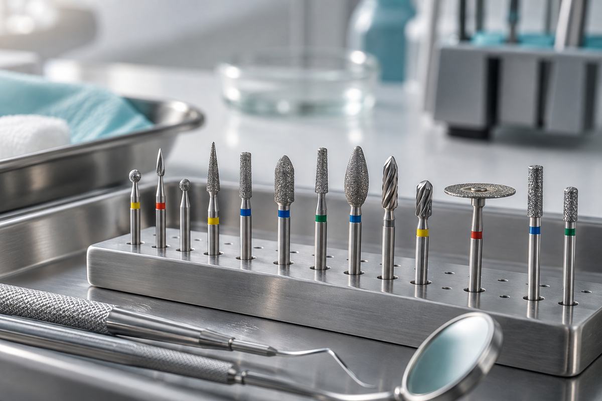
For clinicians working with zirconia-based restorations over implants, selecting the right finishing instruments is equally important. High-quality diamond dental burs ensure precise adjustments to the final prosthesis without damaging the implant components.
What These Trends Mean for Your Practice
The convergence of better materials, surface science, digital planning, and robotic assistance is making dental implant therapy more predictable and accessible than ever. While not every practice will adopt robotic surgery immediately, the shift toward digital workflows and evidence-based surface technologies is already well underway.
For practitioners looking to stay current, investing in digital planning software and understanding the science behind modern implant surfaces will deliver the most immediate returns. For patients, these advances translate to shorter treatment times, fewer complications, and restorations that function reliably for decades.
To learn more about how technology is shaping modern implant practice, read our article on the future of dental implants and how technology is changing tooth replacement.