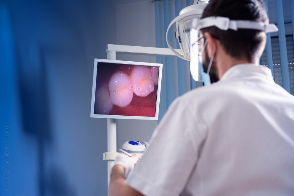
The Evolution of Intraoral Camera Technology
Intraoral cameras have fundamentally changed how dentists communicate findings and how patients understand their oral health. These compact imaging devices capture high-resolution images and video inside the mouth, displaying conditions on chairside monitors in real-time. The technology bridges the gap between clinical observation and patient comprehension, transforming abstract diagnoses into visual evidence.
Early intraoral cameras appeared in the 1980s with bulky designs and poor image quality. Modern systems feature miniaturized sensors, LED illumination, and wireless connectivity. Image resolution now rivals professional photography, capturing detail invisible to the naked eye. This technological advancement enables earlier diagnosis and more predictable treatment outcomes.
Types of Intraoral Cameras for Dental Practices
Wired Intraoral Cameras
Wired cameras connect directly to computer systems via USB interfaces. These devices offer consistent power supply eliminating battery concerns during clinical use. Image transmission occurs instantaneously without wireless lag or connectivity issues. Wired systems typically cost less than wireless alternatives while providing comparable image quality.
The primary limitation involves cable management during patient examination. Proper technique requires positioning the cable to avoid contamination and maintain ergonomic hand positions. Many practitioners prefer wired cameras for fixed operatory setups where cable routing integrates cleanly into the workspace design.
Wireless Intraoral Cameras
Wireless cameras transmit images via WiFi or proprietary radio frequencies to receiving stations. Freedom from cables improves maneuverability and reduces cross-contamination concerns. Battery-powered operation allows use across multiple treatment rooms without additional equipment installation.
Modern lithium batteries provide 2-4 hours of continuous operation between charges. Wireless systems cost more initially but offer superior flexibility for practices with multiple operatories or mobile dental services. Image compression for wireless transmission may slightly reduce quality compared to wired connections, though advances minimize this difference.
Integrated Camera Systems
High-end dental units incorporate cameras directly into handpiece systems or mounted on articulating arms. Integration with chair controls and foot pedals simplifies image capture during procedures. Automated positioning systems maintain consistent camera angles for standardized documentation.
These systems represent significant capital investment but provide seamless workflow integration. Shared databases automatically link captured images to patient records. Multi-monitor setups display images simultaneously to patients and clinical staff, enhancing communication efficiency.
Essential Features in Modern Intraoral Cameras
| Feature | Clinical Benefit | Recommended Specifications |
|---|---|---|
| Image Resolution | Detail visibility for accurate diagnosis | Minimum 1080p, 4K preferred |
| LED Illumination | Consistent lighting without heat generation | Adjustable intensity, 5000-6500K color temperature |
| Autofocus System | Sharp images at varying distances | Continuous autofocus, 5-50mm focal range |
| Video Capability | Dynamic recording of procedures | 30-60 fps, HD or 4K resolution |
| Software Integration | Seamless patient record documentation | Compatible with major practice management systems |
| Autoclavable Components | Infection control compliance | Removable camera sheaths or tips |
Resolution and Image Sensor Quality
Sensor size and pixel density determine diagnostic image quality. Full HD (1920x1080) represents the minimum acceptable resolution for clinical documentation. Ultra HD 4K sensors (3840x2160) capture four times the pixel data, revealing microfractures, early caries, and subtle margin discrepancies invisible at lower resolutions.
CMOS sensors dominate current camera designs due to low power consumption and high-speed image processing. Larger sensors capture more light, improving performance in shadowed areas like subgingival margins. Back-illuminated sensor designs further enhance low-light sensitivity without increasing image noise.
Illumination Systems and Color Accuracy
LED arrays surrounding the camera lens provide shadow-free illumination at all focal distances. Color temperature around 5500-6500K (daylight spectrum) ensures accurate shade matching and tissue color representation. Adjustable intensity prevents overexposure when imaging reflective surfaces like polished restorations.
Polarizing filters reduce glare from wet tooth surfaces and metallic restorations. This optical enhancement improves margin visibility and surface texture documentation. Some advanced systems offer switchable polarization modes for different clinical applications.
Connectivity and Software Integration
Modern cameras connect via USB 3.0 for fast data transfer or WiFi for wireless operation. Compatibility with practice management software eliminates manual image importing. Direct capture buttons in patient records simplify documentation workflow, reducing chair time.
Cloud-based image storage enables remote consultation with specialists and lab communication. HIPAA-compliant encryption protects patient data during transmission and storage. Automated backup systems prevent image loss from hardware failures.
Clinical Applications Transforming Patient Care
Enhanced Diagnostic Capabilities
Magnified intraoral imaging reveals conditions in early developmental stages. Incipient caries appear as opacity changes before cavitation occurs. Marginal gaps in existing restorations measure 50-100 microns become visible. Crack lines in teeth predict future fracture patterns allowing preventive intervention.
Periodontal documentation captures calculus deposits, gingival inflammation, and recession patterns. Sequential images track disease progression or healing response objectively. Comparison tools overlay historical images revealing subtle changes over time.
Patient Education and Treatment Acceptance
Visual evidence transforms patient understanding compared to verbal descriptions alone. Studies demonstrate treatment acceptance rates increase 40-60% when patients view intraoral images versus mirror examination only. Fear and skepticism reduce when patients see actual conditions requiring treatment.
Side-by-side comparisons show healthy versus diseased tissue states. Annotation tools highlight specific problems with arrows and text labels. Before-and-after documentation demonstrates treatment success, building confidence in recommended care.
Documentation for Insurance and Legal Protection
Detailed photographic documentation supports insurance claims with objective evidence of treatment necessity. Pre-operative images establish baseline conditions. Post-operative documentation confirms treatment completion and quality.
complete records provide legal protection in malpractice situations. Time-stamped images create indisputable treatment timelines. Standardized documentation protocols ensure consistent record quality across all patient encounters.
Specialist Referrals and Lab Communication
High-resolution images attached to referral communications give specialists detailed information before patient arrival. Endodontists evaluate crack patterns and pulp chamber anatomy. Periodontists assess soft tissue architecture and recession severity. This advance preparation improves consultation efficiency and treatment planning accuracy.
Laboratory technicians benefit from color-accurate images showing adjacent teeth for shade matching. Preparation margin documentation ensures restoration fit. Direct visual communication reduces remake rates and improves final restoration aesthetics.
Integration with Practice Management Software
Seamless integration with existing practice systems maximizes intraoral camera value. Leading practice management platforms include native camera support with one-click capture into patient charts. Images automatically link to specific tooth numbers and procedure codes simplifying documentation.
Treatment planning modules display captured images alongside proposed procedures. Financial presentation software incorporates before images into patient payment plans. Marketing automation systems use selected images for patient education emails and social media content with proper consent.
Workflow Optimization Strategies
Efficient camera implementation requires protocol development. Standardized image series for new patient exams ensure consistent documentation. Specific views for different procedures create repeatable workflows reducing decision fatigue.
Voice-activated capture allows hands-free operation maintaining sterile fields. Foot pedal controls integrate with dental unit systems. Dedicated monitors positioned for patient visibility eliminate the need for screen sharing or patient repositioning.
Selection Criteria for Practice Needs
Choosing appropriate intraoral camera systems requires evaluating multiple factors beyond initial purchase price:
- Practice Size: Solo practices benefit from single wired cameras, while multi-doctor offices require wireless systems for operatory sharing
- Patient Demographics: Pediatric practices need rugged, easily cleaned designs; cosmetic practices require 4K resolution for detailed documentation
- Existing Technology: Compatibility with current practice management software and computer systems prevents integration costs
- Clinical Focus: Restorative practices emphasize color accuracy; periodontal practices need excellent low-light performance
- Budget Constraints: Entry-level cameras start around $1,500; premium systems with advanced features cost $5,000-$8,000
- Warranty and Support: Minimum 2-year warranties with responsive technical support prevent productivity losses from equipment failures
Evaluating Camera Ergonomics
Physical camera design affects clinical usability. Balanced weight distribution reduces hand fatigue during extended use. Textured grips maintain control with gloved hands. Single-handed operation allows simultaneous tissue retraction improving image access.
Camera head size determines posterior access capability. Smaller profiles (12-15mm diameter) reach distal surfaces of second molars. Angled heads improve visibility in difficult areas compared to straight designs. Rotating lens systems eliminate the need for camera repositioning during exams.
Return on Investment Analysis
Intraoral cameras generate positive financial returns through multiple revenue channels. Treatment acceptance improvement represents the primary benefit. Practices report 30-50% increases in same-day treatment acceptance when presenting visual evidence.
Case value increases as patients understand complete treatment needs rather than accepting minimal interventions. Insurance reimbursement improves with photographic documentation supporting medical necessity. Reduced chair time from efficient communication compensates for equipment costs over 12-18 months.
Calculating Practice-Specific ROI
A practice producing $800,000 annually with 20% treatment acceptance can project outcomes from camera implementation. Increasing acceptance to 30% generates $400,000 additional production annually. Even conservative 25% acceptance yields $200,000 increase. With $5,000 camera investment, payback occurs in 1-2 weeks of additional production.
Indirect benefits include reduced failed appointments from improved patient understanding, decreased emergency visits through preventive care acceptance, and enhanced practice reputation from modern technology adoption. Patient referrals increase when existing patients experience clear communication and feel involved in treatment decisions.
Maintenance and Longevity Considerations
Proper maintenance extends camera lifespan to 7-10 years of clinical service. Disposable sheath systems eliminate direct contamination requiring only external wipe-down between patients. Autoclavable tip designs require careful handling preventing o-ring damage and seal failures.
Regular lens cleaning maintains image clarity. Microfiber cloths remove debris without scratching optical surfaces. Alcohol-based solutions clean external housings between uses. Avoid immersion cleaning unless specifically approved by manufacturers.
Software updates improve functionality and maintain compatibility with evolving practice management systems. Firmware updates enhance autofocus performance and add new features. Regular backup of image databases prevents data loss from hardware failures.
Training Staff for Maximum Utilization
complete team training ensures consistent camera use across all appointments. Hygienists capture periodontal documentation during cleanings. Assistants document pre-operative conditions and post-operative results. Dentists focus on diagnostic imaging and patient consultation.
Standardized protocols define which images to capture for different appointment types. New patient exams include full mouth series from standard angles. Emergency visits document chief complaint areas. Restorative appointments capture preparations and final restorations.
Role-playing exercises develop comfort discussing images with patients. Team members practice pointing out conditions without alarming patients. Positive framing emphasizes solutions rather than problems, maintaining patient confidence while accepting necessary treatment.
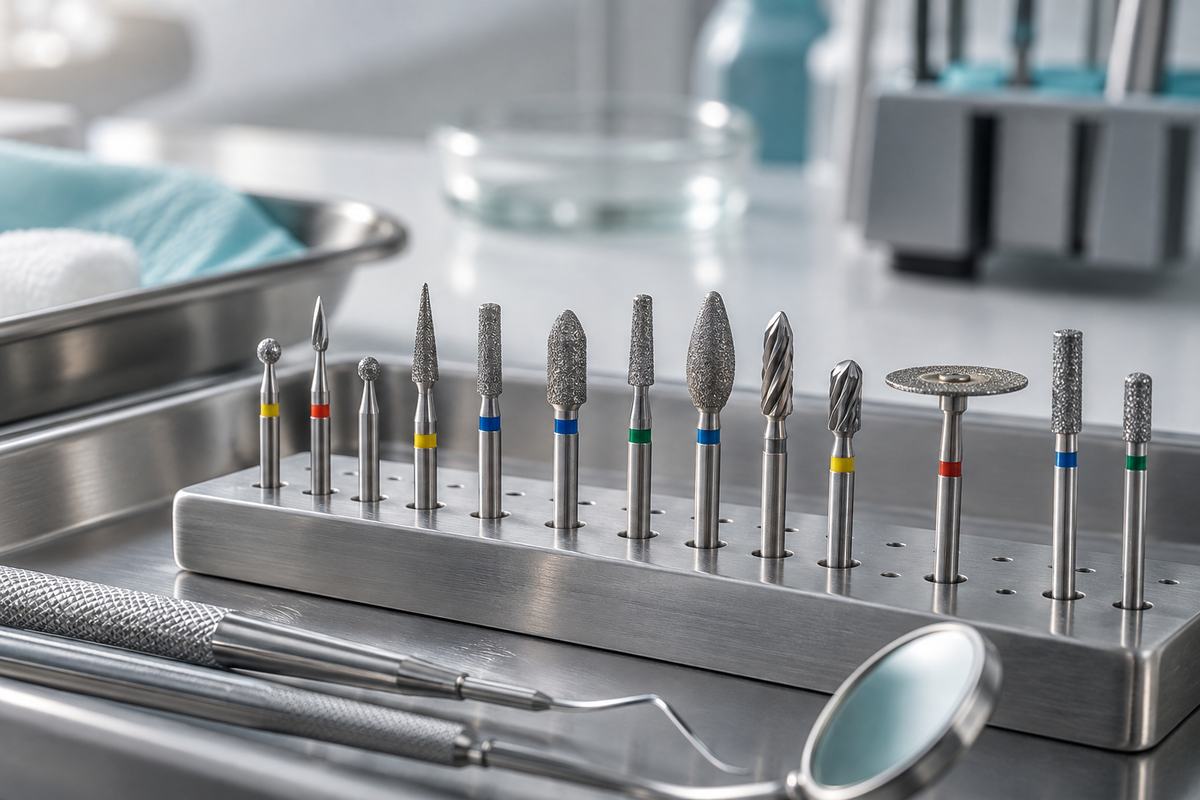
When preparing teeth and restorations captured with your intraoral camera, precision instruments matter. Explore our diamond dental burs for smooth cavity preparation and our silicone rubber polishers for achieving excellent final restoration aesthetics that photograph beautifully.
For more information on dental imaging technology, read our complete guide on benefits of digital dental x-ray sensors and learn about choosing the right dental intraoral camera for you.